
Which types of electron microscopes are there?
One can distinguish three main types of electron microscopes
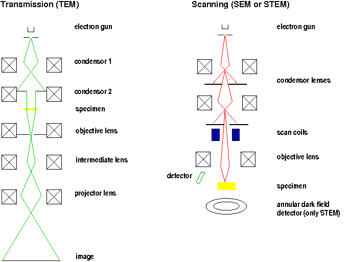
- the scanning electron microscope (SEM)
- The image is formed by scanning a focussed electron beam over the specimen, which can be a bulk one.
- the transmission electron microscope (TEM)
- The beam paths are analogous to those in an optical microscope. Very thin specimens are needed.
- the scanning transmission electron microscope (STEM)
- The construction is the same as in a TEM, but an additional scanning device makes it possible to scan the beam. The pictures have a resolution as high as in a TEM but are recorded in a serial fashion.
SEM
Scanning electron microscopes are used for the investigation of bulk specimens, so there is no need for complex specimen preparations. The acceleration voltage ranges from 1-30 kV. In order to avoid charge effects the specimen should be coated by a metal. An electron probe is generated by demagnifying the crossover into a diameter of about 1-10 nm and scanned in a raster over the specimen.
The picture is generated by synchronizing a cathode ray tube (CRT) with the signal arising from scanning. Either the secondary electrons (SE) or the backscattered electrons (BSE) are detected and deliver information about the topography, the crystal orientation, the material or even magnetic/voltage contrast. Besides, the emitted x-rays or Auger electrons can be sent through a spectrometer to characterize the material.
TEM, STEM
Applying the transmission mode necessitates very thin specimen (5-100 nm for 100 kV). The acceleration voltage is usually in the range between 100-200 kV. Higher voltages up to 300 kV deliver better transmission and higher resolution, but necessitate large constructional systems.
TEM, SEM, STEM
The ray paths are analogous to the scheme of a light microscope (condenser illumination of the specimen). A TEM can also be used in a scanning mode (STEM). For this application an electron probe is formed and scanned over the specimen, so that position dependant information can be extracted.
Depending on whether the intermediate lenses focus the image or the diffraction plane, either the transmission image or the diffraction image is magnified.
The following picture shows a sketch of the respective beam paths (according to the recommendable textbook "Transmission Electron Microscopy" by Ludwig Reimer and Helmut Kohl).
Here you can learn more about the different components of electron microscopes.